Services
Ocular Coherence Tomography
Service Information
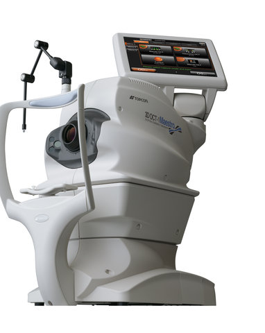
Ocular Coherence Tomography (OCT)
Optical coherence tomography (OCT) is an imaging test that provides detailed images of the optic nerve and layers of the retina. This imaging technique uses coherent light to capture two- and three-dimensional images of the back of the eye with micrometer-resolution. Once processed, eye care professionals are provided with detailed cross-sectional images depicting the different structures in the eye. OCT is a simple and non-invasive test that takes only a few minutes to complete.
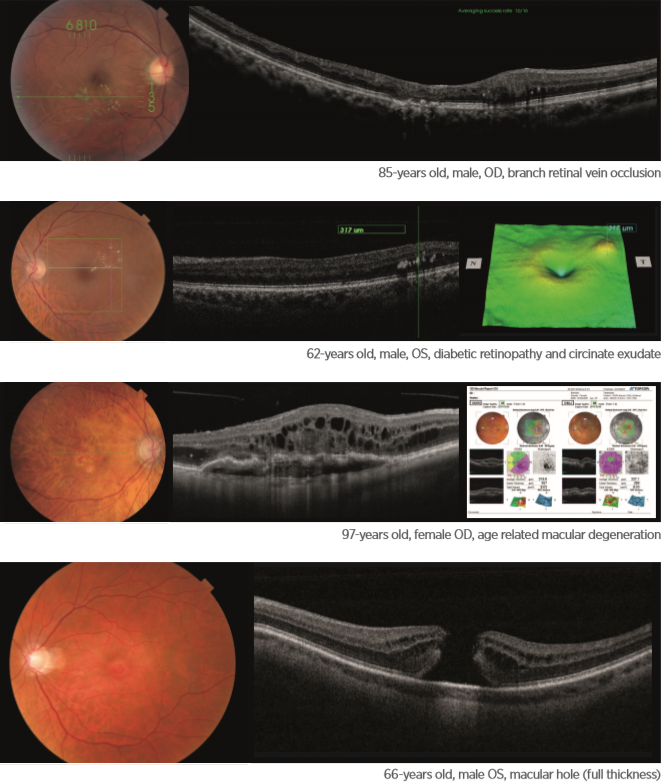
Clinicians are now using OCT in clinical practice for both anterior (front of the eye) and posterior (back of the eye) segment pathologies, as it provides valuable data that can aid in the detection of ocular pathologies, as well as track progression of the condition and the response to treatment. For example, OCT can help detect optic neuropathies with retinal nerve fiber layer (RNFL) loss, such as in glaucomatous damage. The instrument can also be used to identify disc edema and even buried disc drusen. Analysis of retinal thickness over the macula and posterior pole can help detect retinal edema or atrophy. Anterior segment OCT can provide further insight into anterior chamber depth, angle anatomy and corneal pathologies.