Services
Glaucoma Testing

Glaucoma
Glaucoma is a group of eye diseases which result in damage to the optic nerve and vision loss. Early detection, through regular and complete eye exams, is the key to protecting your vision from damage caused by glaucoma. If the doctor suspects glaucoma, there are a variety of tests to help diagnose, monitor and control glaucoma.
- Intraocular Pressure
- Gonioscopy
- HRT III
- OCT
- Pachymetry
- Visual Field
Measuring the pressure in the eye has long been a standard of care in the diagnosis of glaucoma. However, there are cases of glaucoma where the intraocular pressure is not elevated (normal tension glaucoma). Therefore, a diagnosis can not be determined by pressure alone. Instead, a combination of pressure readings, corneal thickness measurements, anterior chamber angle evaluation, and optic nerve and visual field analysis is required to accurately diagnose all cases of glaucoma.
This test involves the use of a special mirrored lens which allows the doctor to examine the anterior chamber angle structures. The doctor can then determine if the eye's drainage route is wide open, narrow or closed, or clogged with pigment or debris. With this information, the doctor has more insight into the type of glaucoma present.
Glaucoma is a progressive disease of the optic nerve that leads to irreversible visual loss. However, clinical studies have confirmed that early detection and treatment of glaucoma can slow the progression of permanent optic nerve damage and blindness.
Common conventional methods of evaluating the optic nerve are very subjective: Doctors try to assess the nerve by way of diagrams to identify glaucomatous cupping and damage. These methods do not provide exact quantitative measurements, and comparisons for subtle changes over time are very difficult. Regardless of their limitations, these methods are considered the current standard of care, and are covered by OHIP.
Now patients have a newer, more advanced option: The Heidelberg Retinal Tomograph III (HRT III) has been proven to be superior to all other available optic nerve imaging techniques, and uses the latest in scanning laser technology for early detection and follow-up of glaucoma. This machine is a laser ophthalmoscope, which can precicely measure and analyze the shape of the optic nerve. The HRT III is not a treatment laser, and is not harmful to the eye.
Data from successive HRT III exams is superimposed on the original data (or baseline exam) for a point-by-point comparison to identify changes over time. Therefore, repeated measurements of the optic nerve using the HRT III can pick up glaucoma damage up to four years earlier than visual field testing. These subtle changes will help to determine when treatment is necessary for glaucoma suspects, or when treatment should be increased or surgery considered for patients with existing glaucoma damage.
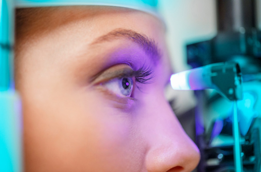
The test is safe and takes moments to complete: Patients look at a target light and must not blink or move the eye for several seconds while the photograph of their optic nerve is being taken. (Dilating drops may be necessary if a patient has cataracts or corneal scarring). The HRT III exam takes place in this office, and the test is used to monitor the progression of your glaucoma. The frequency of follow-up exams is determined by your doctor, and based on whether the progression of your glaucoma is stable or aggressive.
The cost of this examination is not covered by OHIP. The examination fee is billed directly to the patient, and a receipt for your health insurance or tax purposes will be issued. Methods of acceptable payment include Interac, Visa, Mastercard, cash, or cheque.
Optical coherence tomography (OCT) is an imaging test that provides detailed images of the optic nerve and layers of the retina. This imaging technique uses coherent light to capture two- and three-dimensional images of the back of the eye with micrometer-resolution. Once processed, eye care professionals are provided with detailed cross-sectional images depicting the different structures in the eye. OCT is a simple and non-invasive test that takes only a few minutes to complete.
Clinicians are now using OCT in clinical practice for both anterior (front of the eye) and posterior (back of the eye) segment pathologies, as it provides valuable data that can aid in the detection of ocular pathologies, as well as track progression of the condition and the response to treatment. For example, OCT can help detect optic neuropathies with retinal nerve fiber layer (RNFL) loss, such as in glaucomatous damage. The instrument can also be used to identify disc edema and even buried disc drusen. Analysis of retinal thickness over the macula and posterior pole can help detect retinal edema or atrophy. Anterior segment OCT can provide further insight into anterior chamber depth, angle anatomy and corneal pathologies.
Pachymetry gives the doctor a measure of the thickness of the cornea, which is another helpful tool in the diagnosis of glaucoma. When intraocular pressure is measured by applanation tonometry (the gold standard), a probe is used which measures the force required to flatten a specific area of the cornea (Pressure=Force/Area). Consider a cornea that is a little thicker than normal. With this thickness, more pressure is required to flatten the cornea the same amount, thus the pressure reading is artificially elevated. On the other hand, with thinner corneas, less pressure is needed to flatten the same amount of cornea, resulting in an erroneously lower pressure reading. With the pachymetry results the doctor is able to adjust the intraocular pressures of the eyes leading to a more accurate representation of the eye's actual pressure.
With glaucoma, vision loss usually starts in the periphery and progresses inward if left untreated. This vision loss is usually asymptomatic and therefore most people do not realize that they have glaucoma until significant damage has been done to the nerve fiber layer. With a visual field machine doctors are able to accurately map out your visual field (i.e. what you are seeing and what you are not seeing). Characteristic patterns of vision loss are then analyzed, providing an excellent basis for the diagnosis of glaucoma. With repeat testing, a visual field analysis can show the progression of vision loss as compared to previous results. This can then be used to monitor and treat glaucoma.